When blood flow into a heart chamber is increased, it causes the chamber to fill more. That distention makes the chamber adapt over time. This paper is about what happens to heart growth in the fetus when blood flow into the right side of the heart is increased. We did that by sewing a passageway between the carotid artery and the jugular vein on one side of the neck. All of the blood from that artery was diverted directly back into the fetal heart.
How a fetus grows in utero determines their risk for heart disease in later life. The fetal heart grows rapidly, in ways that are fundamentally different than how the adult heart grows. It is important to understand how increased filling by blood affects fetal heart growth, because this may alter cardiac risk for life.
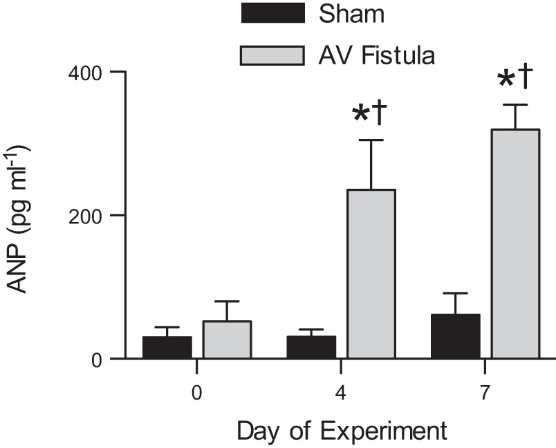
Now we know that when filling of the right side of the fetal heart is increased, the heart muscle cells grow larger. This is a different response than when blood pressure is increased in the fetus, which also causes heart muscle cells to make more of themselves and to mature.
The stimulus of increased filling caused a type of fetal cardiac muscle adaptation that we haven’t seen with other stimuli. Knowing how distention affects fetal heart muscle growth will help us interpret how other conditions, such as low oxygen, change fetal development.
Find this paper the American Journal of Physiology and on PubMed.